Tumors of the RPE > RPE Hypertrophy + CHRPE + Bear Tracks Retina Rocks
The retina is an ocular structure responsible for gathering gross visual stimuli that enter the eye and transforming them into neuronal signals transported to the brain for interpretation. Truly, it comprises two distinct entities that work in concert with each other: the neural retina and the retinal pigmented epithelium.

Bear tracks retina gertyleo
The retinal pigment epithelium (RPE) is a pigmented layer of the retina which can be thicker than normal at birth (congenital) or may thicken later in life. Areas of retinal pigment epithelial (RPE) hypertrophy usually do not cause symptoms. They are typically found during routine eye examinations.
Tumors of the RPE > RPE Hypertrophy + CHRPE + Bear Tracks Retina Rocks
March 9, 2020 at 4:43 p.m. EDT. Trainer Jason Servis is among those charged by federal prosecutors with participating in a widespread doping scheme. (Gregory Payan/AP) More than two dozen horse.

Bear Track / CHRPE // Congential Hypertrophy of Retinal Pigment epithelium // FAP lesions
Congenital hypertrophy of the retinal pigment epithelium (CHRPE) is a generally asymptomatic congenital hamartoma of the retina. Typical (solitary and grouped) and atypical variant forms are described. Atypical CHRPE is associated with familial adenomatous polyposis (FAP). Contents 1Disease Entity 1.1Disease 1.2Epidemiology 1.3Risk Factors
Bear Tracks Retina Image Bank
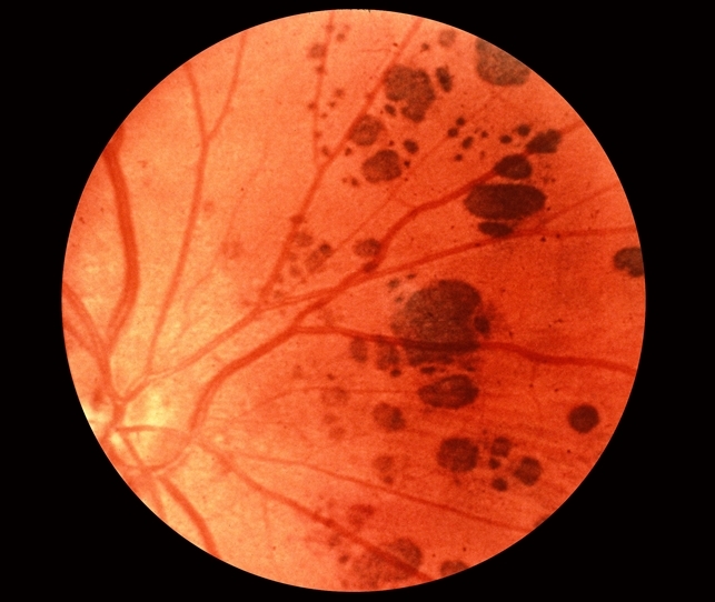
Grouped congenital hypertrophy of the retinal pigment epithelium is a conspicuous ocular anomaly wherein highly pigmented, demarcated but flat retinal lesions arise from the retinal pigment epithelium. These lesions ("bear tracks") typically increase in size as they approach the retinal periphery. The discovery of pigmentary lesions in a young infant with a poor red reflex warrants urgent.

Bear Tracks CHRPE em paciente com história familiar de ad… Flickr
The differential diagnosis for multiple hyperpigmented lesions in the fundus includes choroidal nevi, RPE hyperplasia secondary to previous trauma or inflammation, pigmented lattice degeneration, congenital hypertrophy of the retinal pigment epithelium (CHRPE), multifocal CHRPE ("bear tracks"), malignant melanoma of the choroid, and RPE hamartom.

Bear Tracks Retina Image Bank
Uploaded on Jan 13, 2020. Last modified by Caroline Bozell on Jan 14, 2020.; Rating Appears in Retina and Choroid Condition/keywords bear tracks, amelanotic, pediatic retina, congenital hypertrophy of the retinal pigment epithelium (CHRPE)
Tumors of the RPE > RPE Hypertrophy + CHRPE + Bear Tracks Retina Rocks
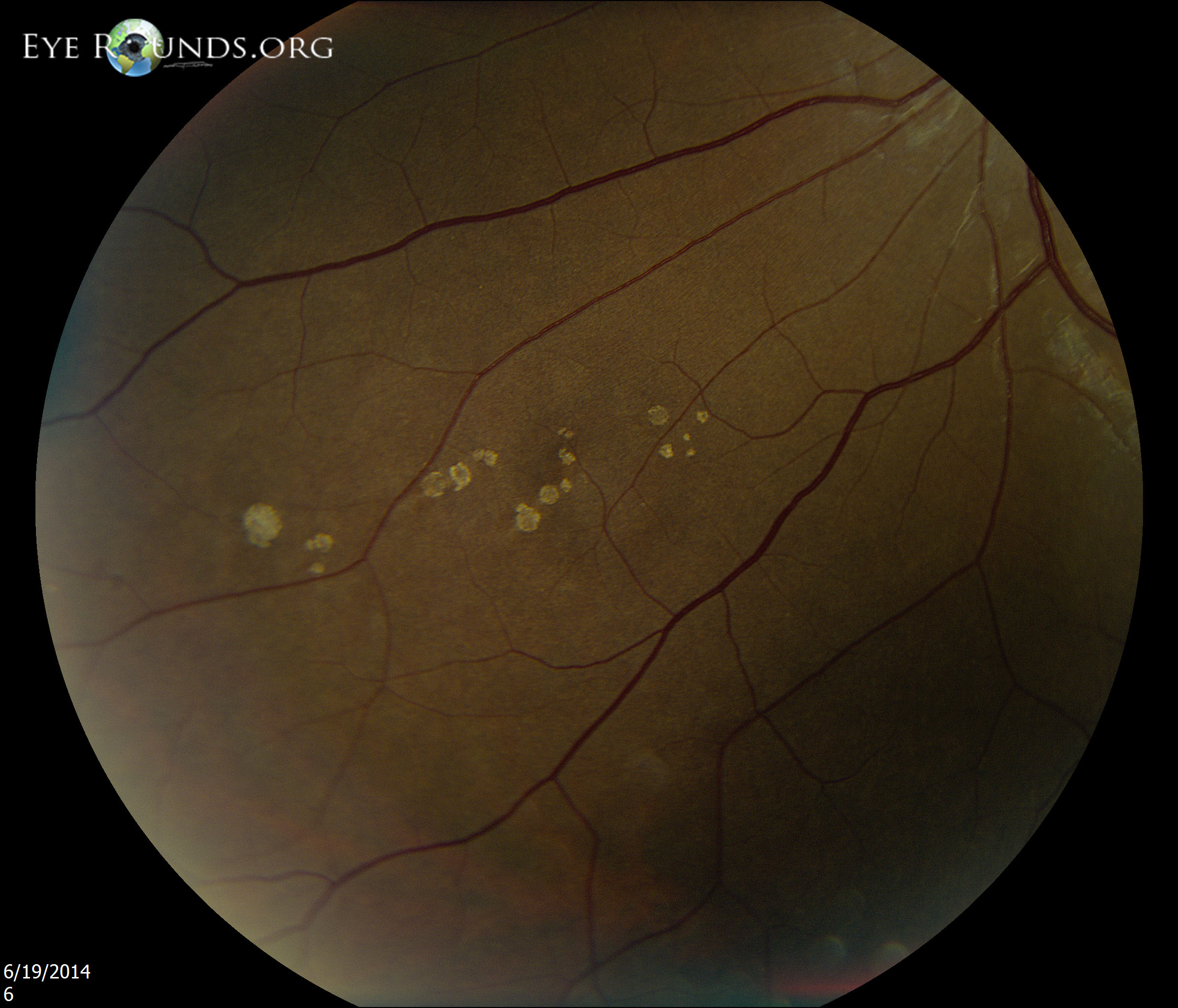
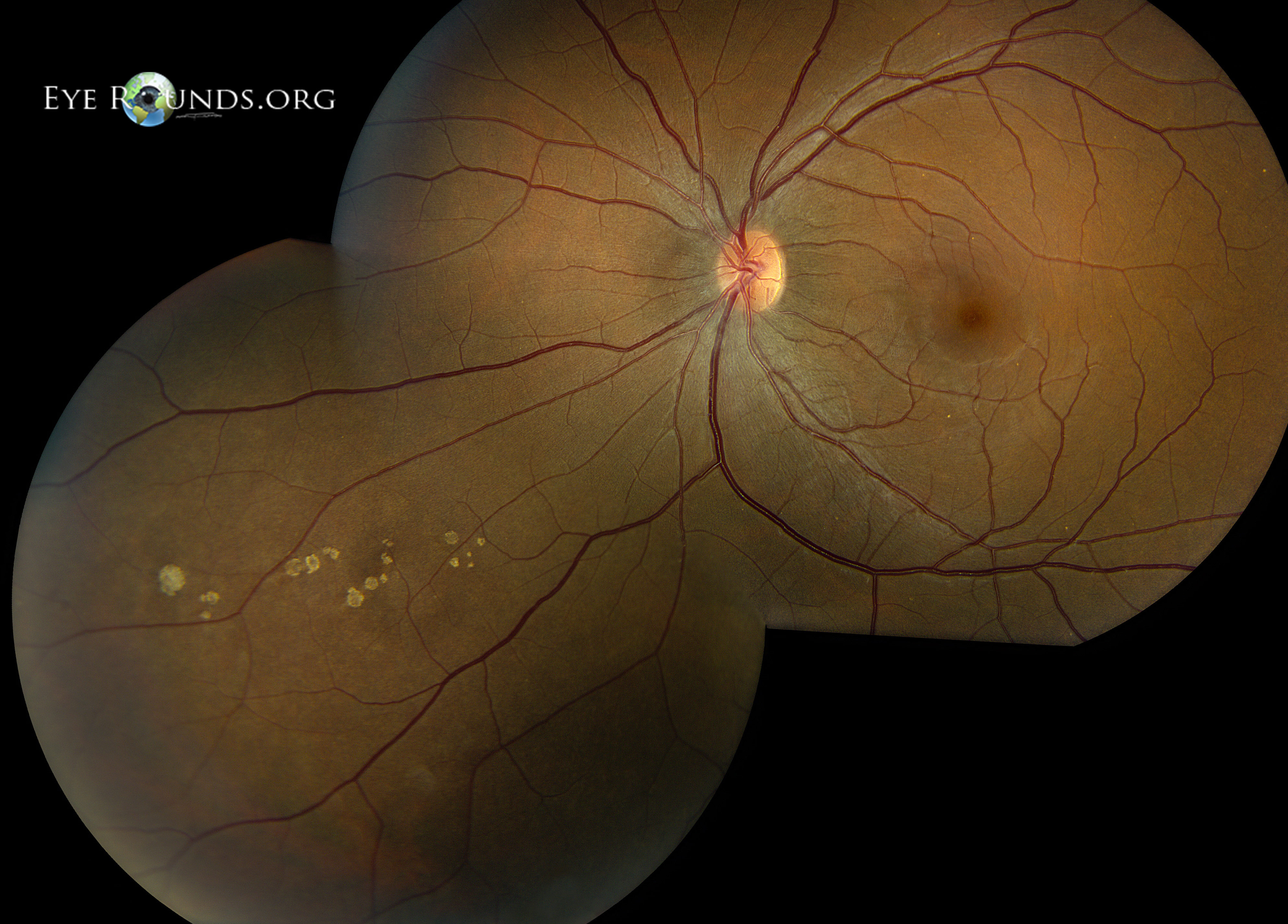
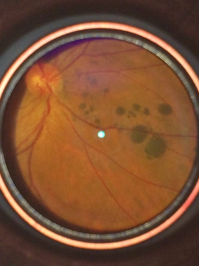
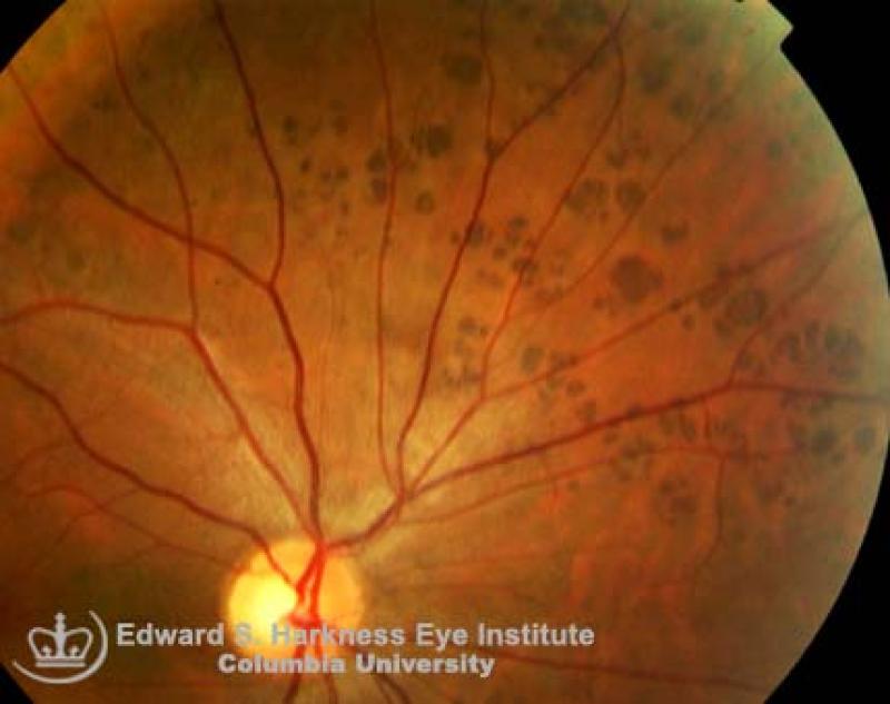
"Bear Tracks" CHRPE Benign pigmented fundus lesions that commonly discovered during routine eye examination. Clinical Features Usually asymptomatic. Signs: Well-demarcated, round, solitary or multiple gray-brown or black lesions which have flat or scalloped margins. May be encircled by hyper- or hypo-pigmented halo.
Atlas Entry Grouped congenital albinotic spots of the retinal pigmented epithelium (polar bear
Diffuse bear-track retina: profound, bilateral, grouped congenital pigmentation of the retinal pigment epithelium in an infant Oliver R. Marmoy, MSc Charlotte Blackwell, Most Sarah Cornelius, BSc Dorothy A. Thompson, PhD Robert H. Henderson, MD Published: October 22, 2020 DOI: https://doi.org/10.1016/j.jaapos.2020.08.003
Bear Tracks Retina Image Bank
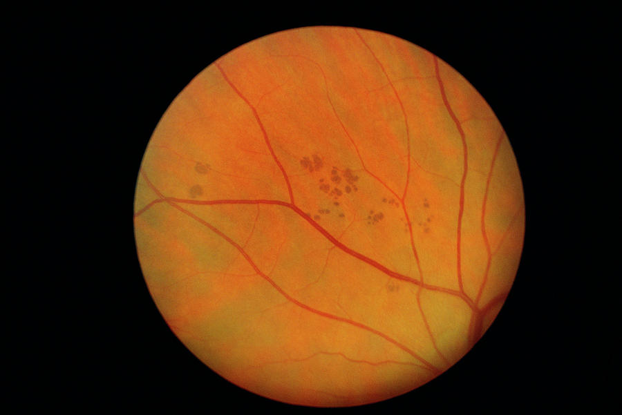
These groupings of round spots of various sizes can resemble paw prints so they are also called Bear Track Pigment. Patients with multiple or irregularly shaped CHRPE are the ones most likely to have FAP and are the ones we educate about the importance of preventive colon health care.

Bear Tracks Pigmentation Retina Image Bank
Congenital hypertrophy of the retinal pigment epithelium is a rare benign tumor of the ocular fundus that may vary according to three types. It is frequently asymptomatic and diagnosed during routine ophthalmology exam.
Atlas Entry Grouped congenital albinotic spots of the retinal pigmented epithelium (polar bear
Diffuse bear-track retina: profound, bilateral, grouped congenital pigmentation of the retinal pigment epithelium in an infant - ScienceDirect Journal of American Association for Pediatric Ophthalmology and Strabismus Volume 24, Issue 6, December 2020, Pages 384-386 Short Report
Bear Tracks On The Retina Photograph by Paul Parker/science Photo Library Pixels
Grouped congenital albinotic spots of the retinal pigment epithelium ('polar bear tracks') A variant of CGP-RPE is congenital albinotic spots of the RPE (CASRPE), also known as 'polar bear tracks'. This condition is characterised by well-circumscribed, chalky white lesions of the same size, shape and distribution as CGP-RPE (Fig. 3a, c)
Bear Tracks Smartphone Fundus Image Retina Image Bank
Congenital hypertrophy of the retinal pigmented epithelium (CHRPE) in a "bear tracks" configuration Category (ies): Genetics, Retina Contributor: Jesse M. Vislisel, MD Posted: August 15, 2013

Name Sara Oelrich C.R.A, C.O.T Description OphthalmicPhotography Retina montage, Bear Tracks
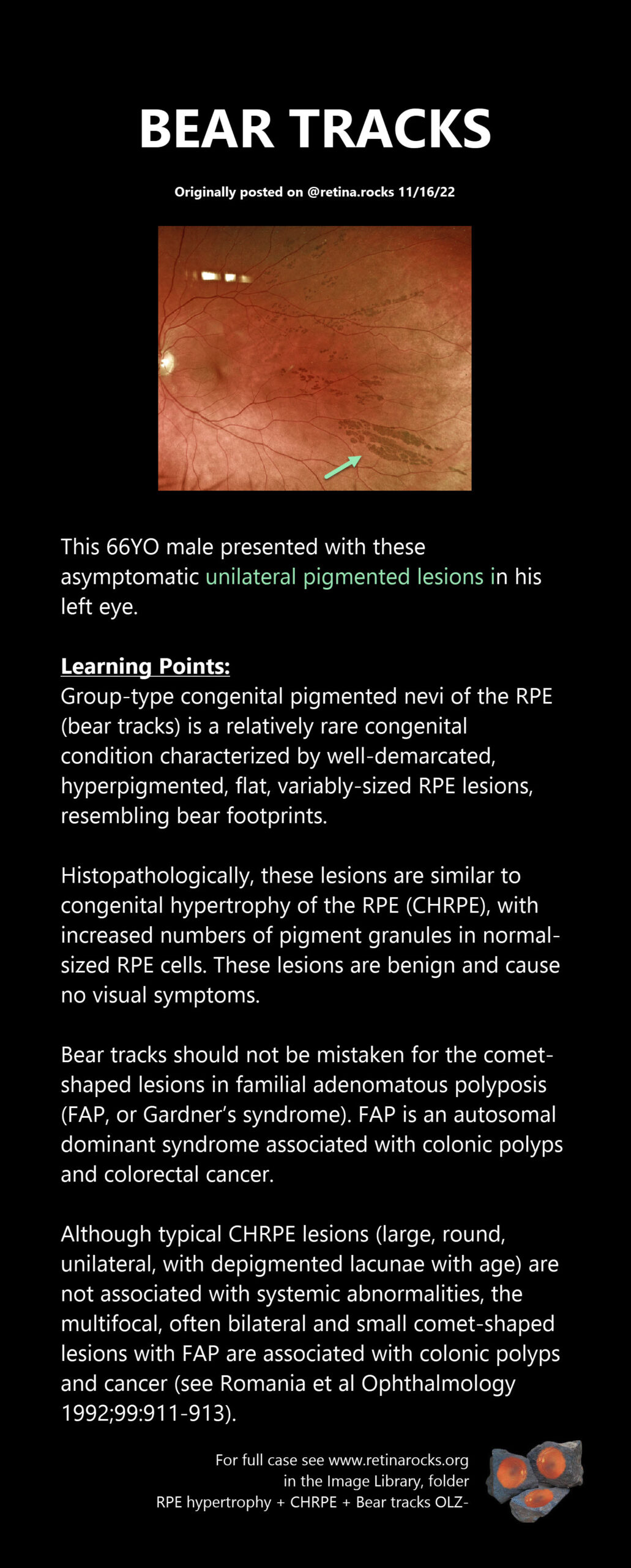
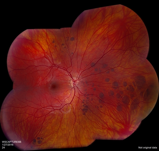
Description. Ultrawide field fundus photograph of a 79-year-old patient who was incidentally found to have extensive bear track lesions in both eyes. Left eye was treated for NVG in the past and bear tracks were only visible temporal to the macula where there was no laser scars. He was referred to be seen by gastroenterologist and have a.
"Bear Tracks" CHRPE Vagelos College of Physicians and Surgeons
Overall, 84% of CHRPE lesions were located in the retinal periphery compared to 16% within the posterior pole. Tourino et al described the most common location for CHRPE amongst FAP individuals was in the retinal equator. 9 92% of FAP individuals had CHRPE present proximal to the retinal vessels compared to 7% in controls (p < 0.001). 9.